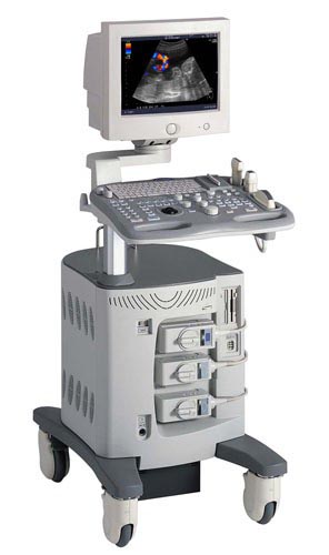
Ultrasound examination (ultrasound) has no contraindications. This is not a radiation method for the study of diseases of adults and children. US - research on expert-class devices allows you to visualize the smallest details, see the smallest structural changes in organs and systems. Ultrasound investigation (ultrasound diagnostics) - examination by means of ultrasonic waves. Ultrasound diagnostics has taken one of the leading places in modern clinical medicine. Dopplerography of blood vessels is the most natural and safe method for a patient to study blood flow in blood vessels. Modern ultrasound technologies make it possible to find the test vessel with high accuracy, assess the nature of blood flow and conduct dopplerometry - a quantitative assessment of blood flow in the area of interest. The use of three-dimensional reconstruction in combination with Doppler mapping allows us to evaluate the architectonics of blood vessels.
Why is ultrasound diagnostics so popular?
- accuracy of the results
- non-invasiveness, atraumatic
- availability, low price
- relative simplicity of the procedure
- can be repeated repeatedly, due to the complete absence of ionizing radiation
Ultrasonic research methods made it possible to more accurately solve the problems of diagnosing a significant number of diseases of the cardiovascular, digestive, genitourinary system, as well as to obtain valuable information in obstetrics and gynecology, oncology. Ultrasound allows you to clearly visualize the vessels and blood flow in them.
We recommend an ultrasound scan to diagnose diseases:
- abdominal organs (liver, gall bladder, pancreas, spleen, kidneys)
- thyroid gland, adrenal gland
- mammary and salivary glands, lymph nodes
- male bladder and prostate
- hearts
- peripheral vessels of the upper and lower extremities (arteries and veins)
- joints and periarticular soft tissues
- examination of the pelvic organs (uterus, ovaries, fallopian tubes and tissues surrounding them)
Ultrasound examination allows you to diagnose a disease even before it has manifested itself clinically, as a result of this treatment can be more timely and effective.
How to prepare for an ultrasound:
Special training require studies of the abdominal cavity and pelvic organs.
Ultrasound scan of the abdomen: 1-2 days before the study, exclude bloating foods (brown bread, legumes, fresh vegetables, fruits, milk, juices, sodas) from the diet, easily have dinner on the eve of the study no later than 19.00 and on an empty stomach see a doctor. If you do not have the opportunity for a morning study, in the morning a light breakfast (for example, unsweetened tea), 6-8 hours before the study.
Ultrasound of the pelvic organs in women: for examination through the abdominal wall (with an abdominal sensor), come with a bladder full, do not empty it for 3-4 hours or drink at least 1 liter of non-carbonated liquid 2 hours before the study
Ultrasound of the bladder and prostate: performed with a full bladder. If you empty the bladder 3-4 hours before the study, then you should drink at least 1 liter of non-carbonated liquid 2 hours before the study.
Ultrasound of the mammary glands is carried out in the first phase (from 7 to 10 days) of the menstrual cycle. Breast ultrasound is a valuable diagnostic addition to mammography.